rahma zakaria
how to grow almost anything
expansion microscopy
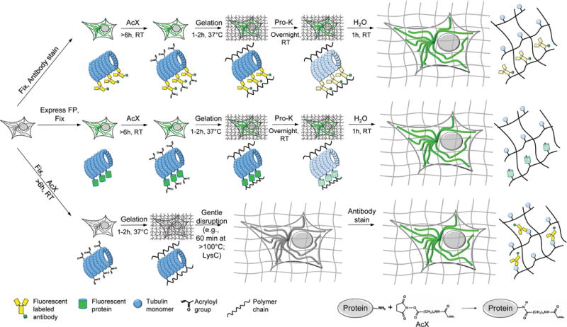
Protein retention expansion microscopy (ProExM) is a powerful technique for expanding microscopic samples. In this technology, proteins in the sample are covalently crosslinked to the gel. When the gel is subsequently expanded/stretched out, the sample also expands. Using this technique, it is possible to image samples beyond the magnification limit of a microscope.
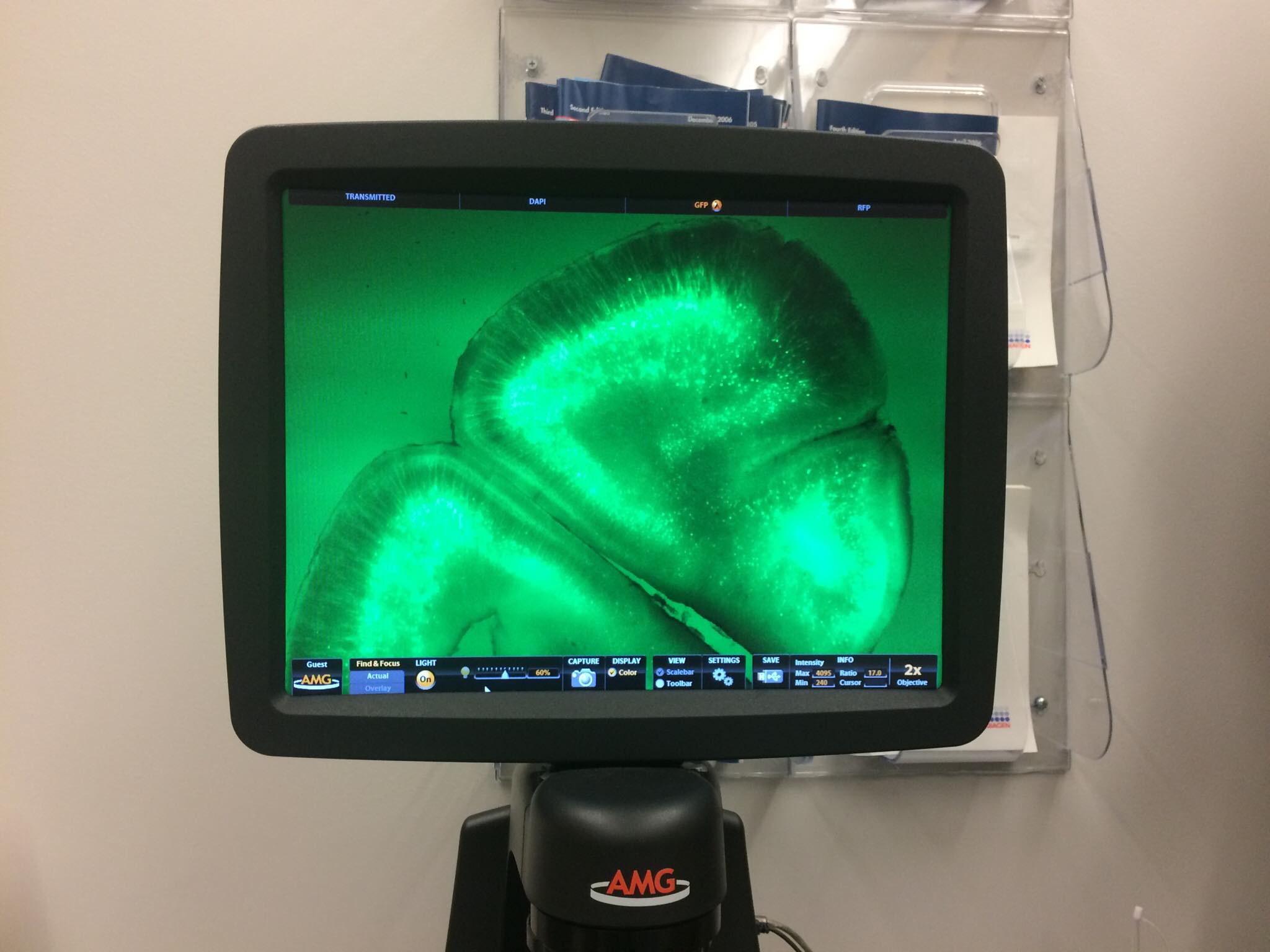
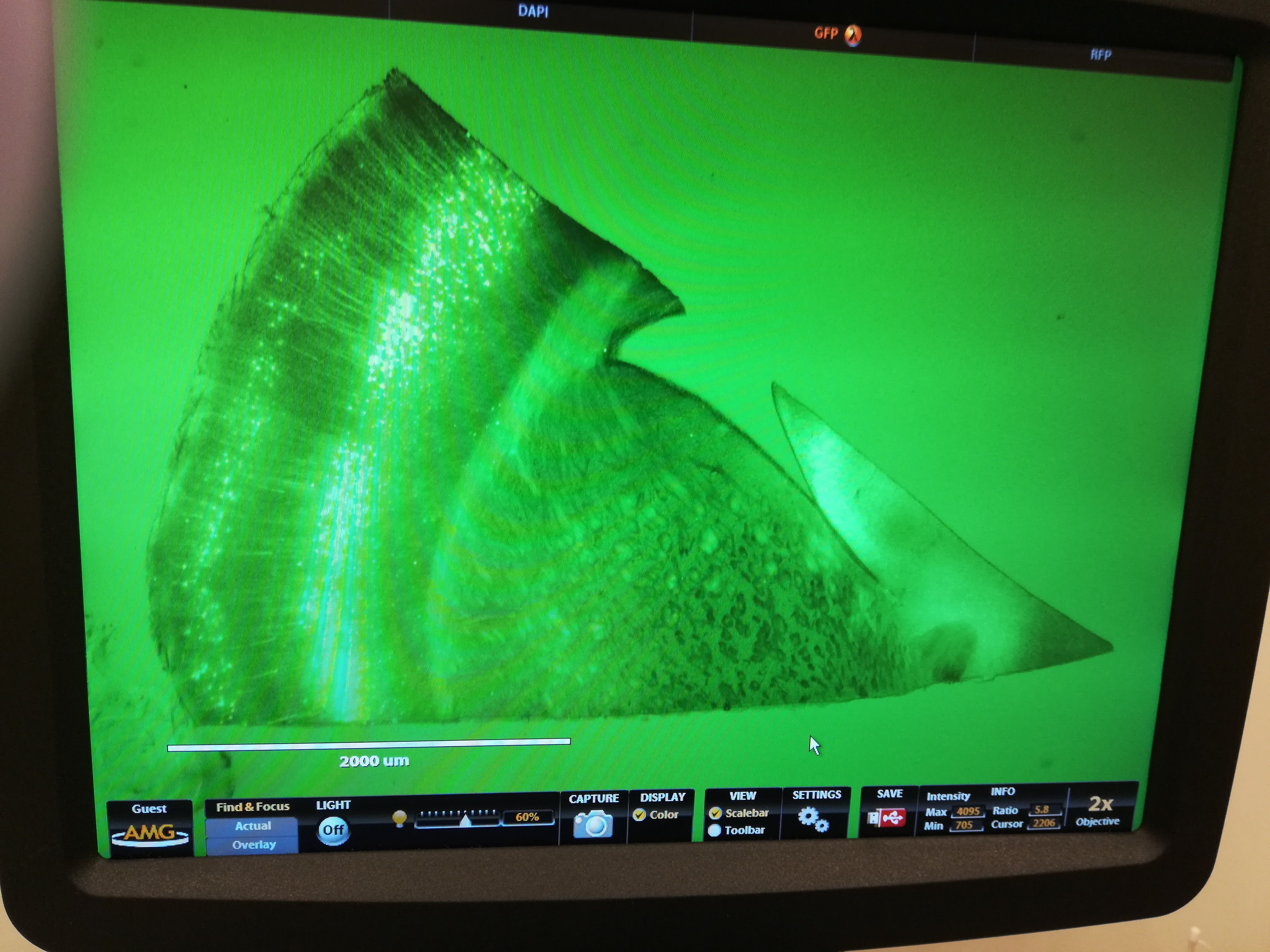
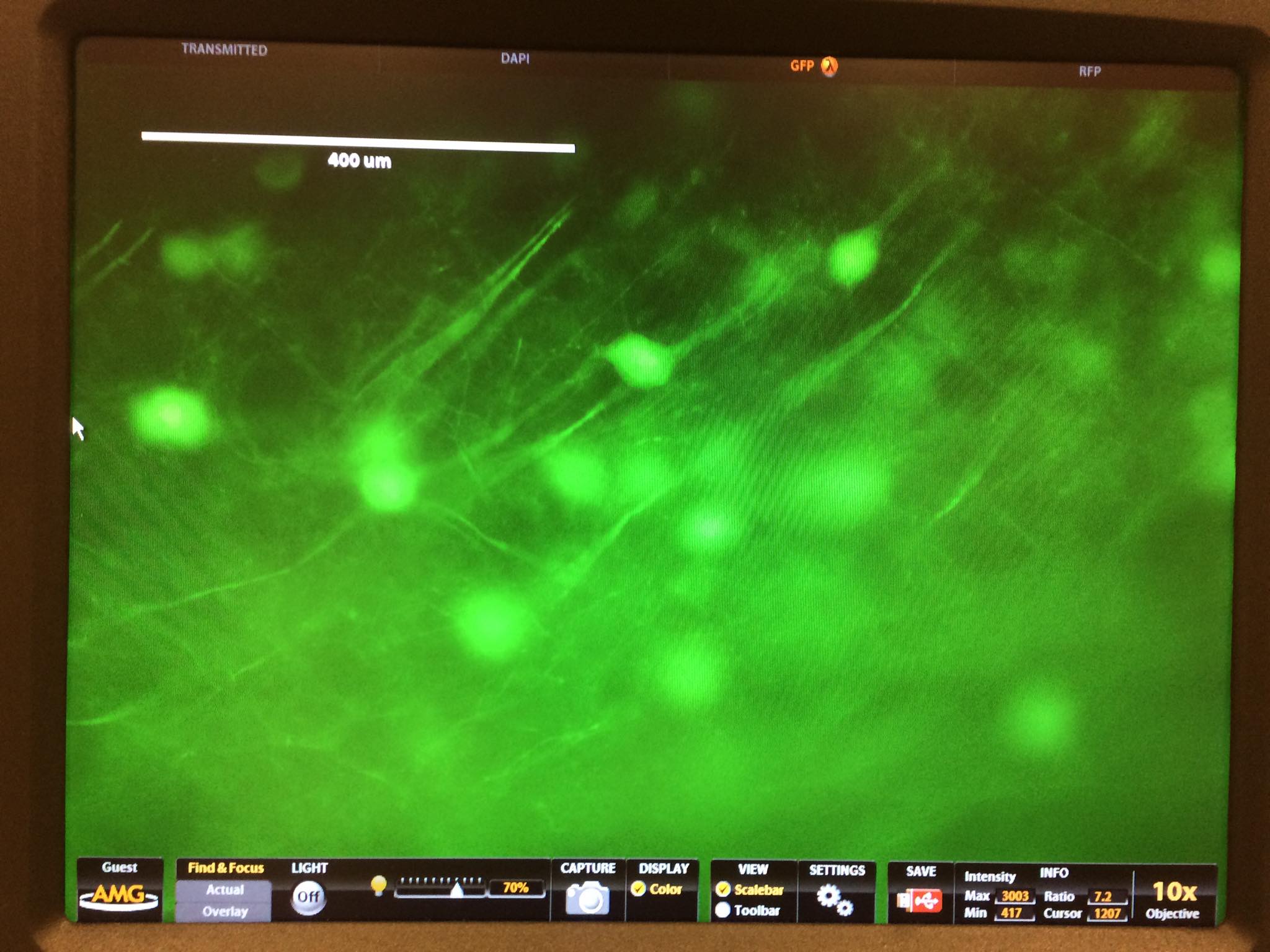
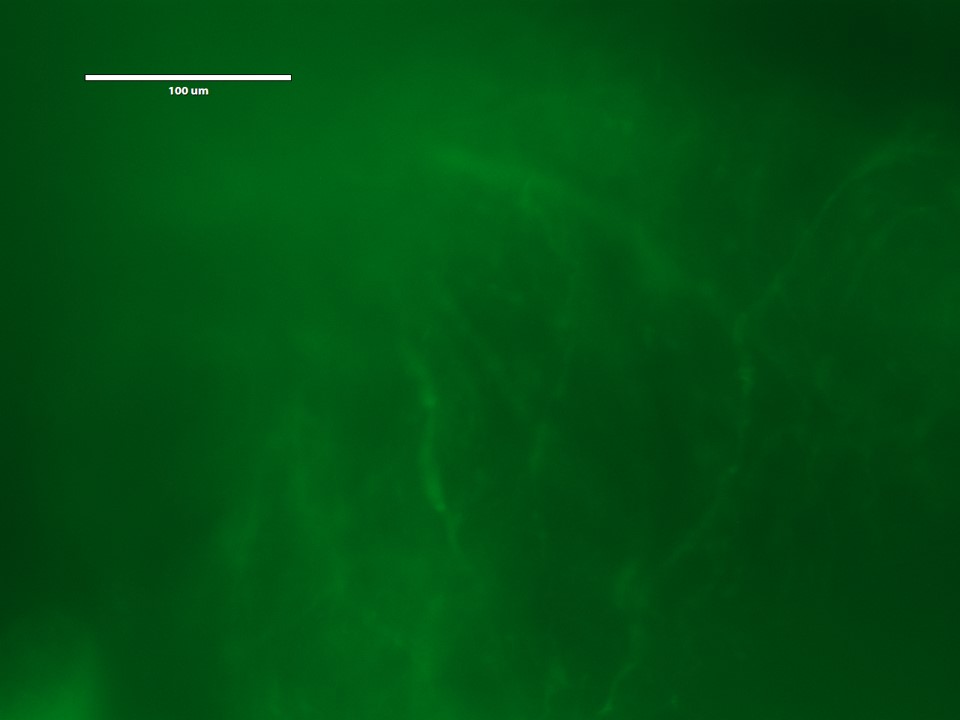
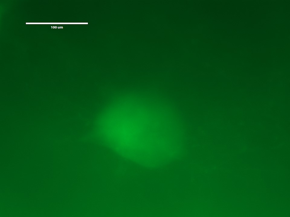
We used ProExM to expand slices of mouse brain tissue, containing neurons expressing YFP.
- (Day 1) Treat brain tissue slice with Acroloyl-X, a molecule which modifies proteins in the sample and enables them to catalyze free-radical polymerization and crosslink to the gel. Image samples.
Acroloyl-X is a toxic molecule!
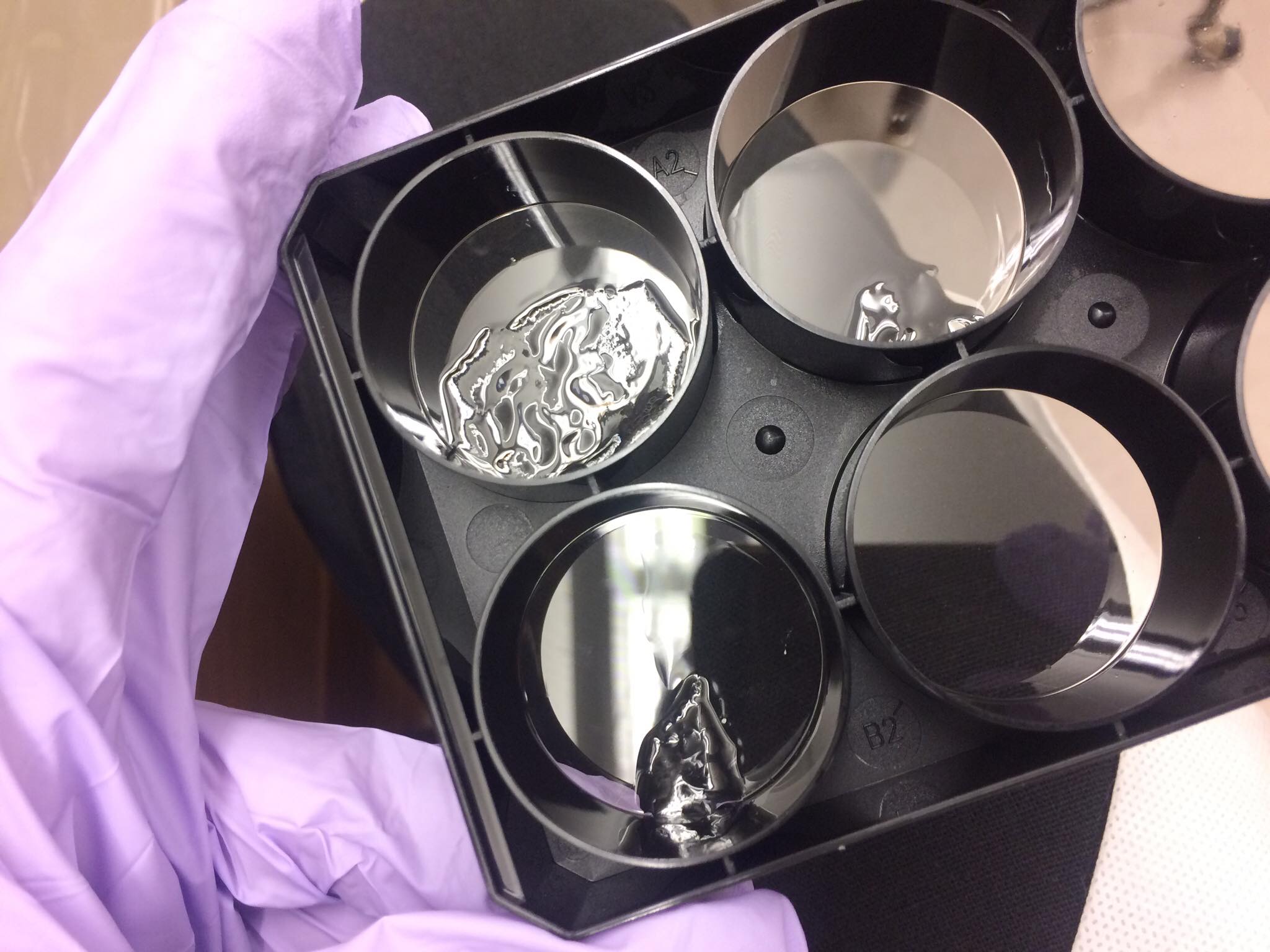
- (Day 2) Embed brain slice in a polyacrylate-polyacrylamide co-polymer gel, such that acryloyl-X-modified proteins in the sample will be incorporated into the gel.
- Incubate gel in a digestion buffer.
- (Day 3) Expand gel through three rounds of washing.
- Image expanded samples
Here are some pictures of our expanded brain slices! We could see the physical expansion of the sample, and under the microscope we could expand with higher resolution.