MAS.S66: How to Grow (Almost) Anything

Priya Pillai

Expansion Microscopy: Using Hydrogels to Enlarge Biological Markers

April 25, 2019

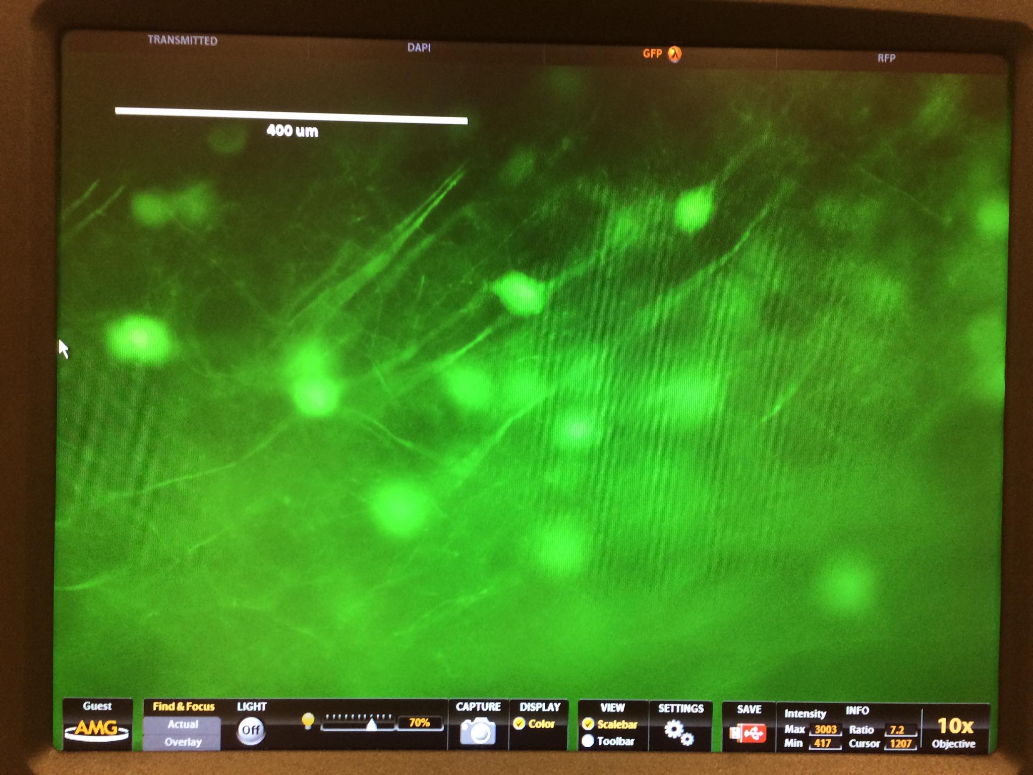
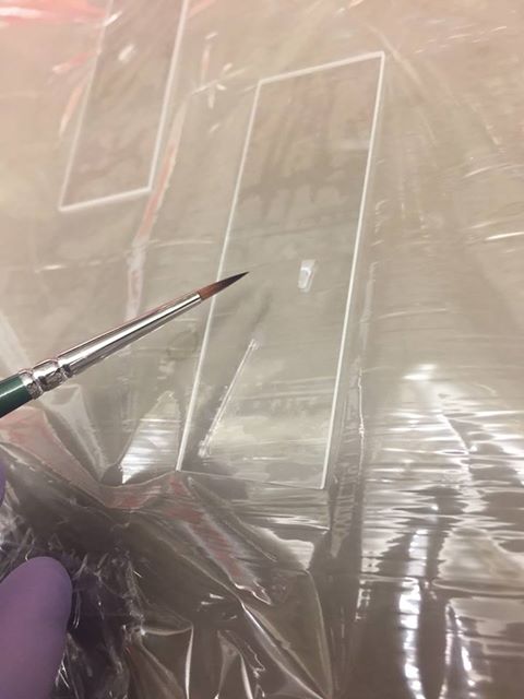
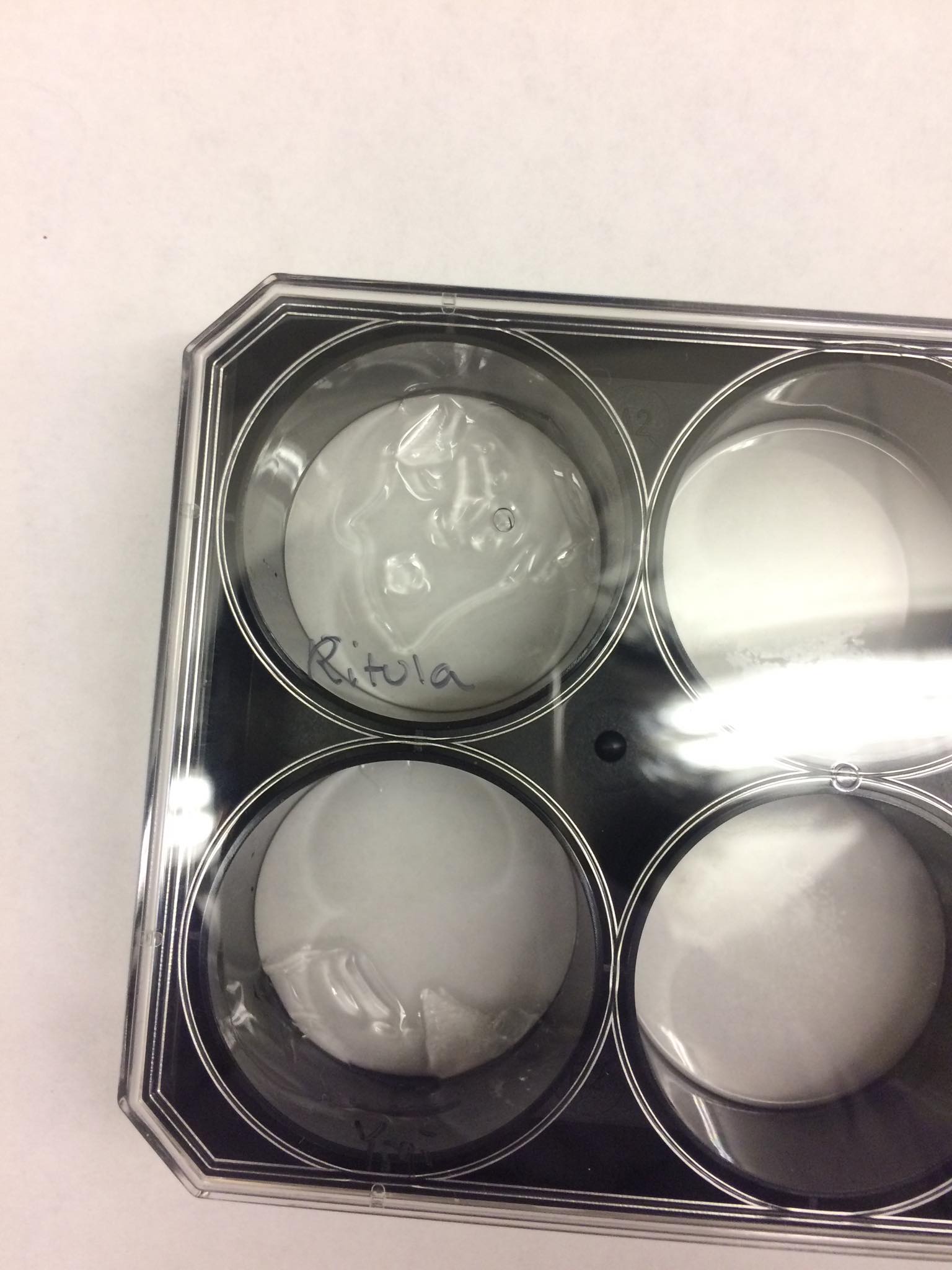
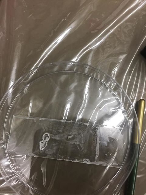
For this experiment, we used Ed Boyden's technique of expansion microscopy to visualize mouse brain neurons labeled with GFP. This works by attaching the GFP markers to a hydrogel in its monomeric state, polymerizing the gel, and then hydrating it to see it expand. We had some issues with expansion due to air bubbles inhibiting polymerization.

Initial Brain Slice Magnified
A fluorscent microscope image of the unexpanded brain slice
Expanded Brain Slice Magnified
A fluorscent microscope image of the expanded brain slice
Initial Brain Slice
The original brain slice
Expanded Brain Slice
The brain slice after it had been expanded with a hydrogel
Dry Brain Slice
A failed expansion due to an air bubble inhibiting hydrogel polymerization