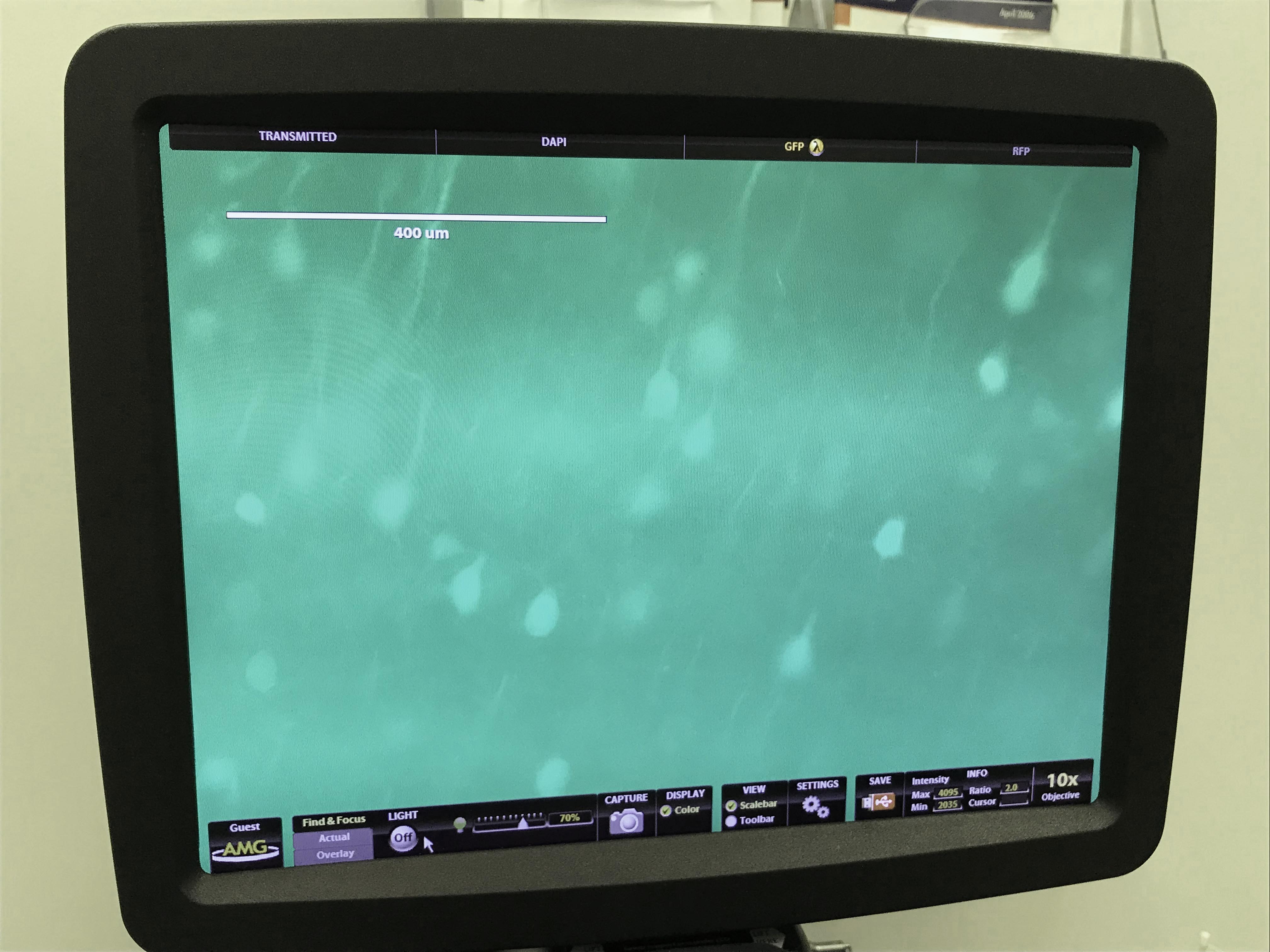
Measurement & Imaging
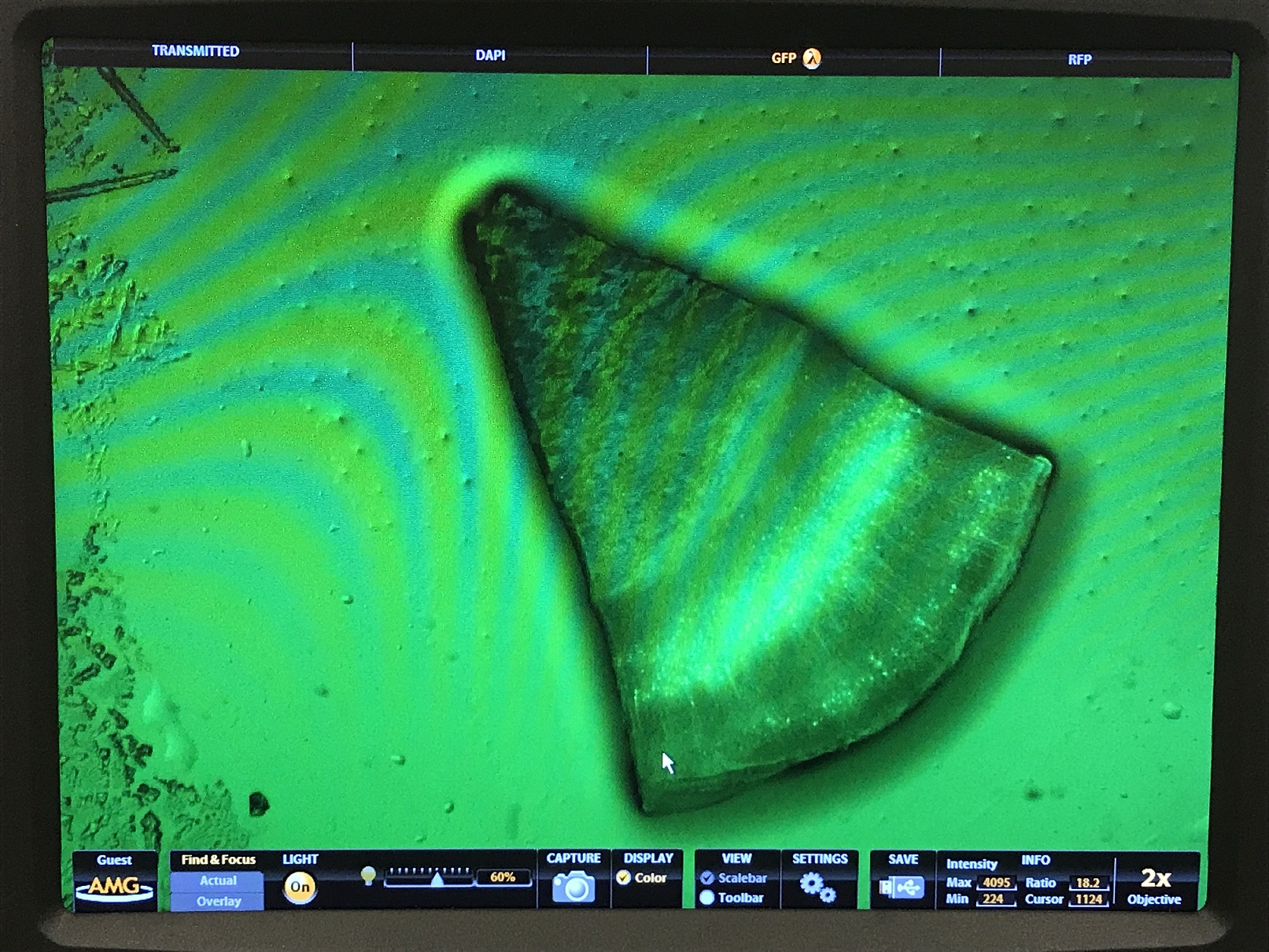
We use Expansion Microscopy (ExM) to to visualize a mouse brain slice with improved resolution on a light microscope. The tissue slices originate from a Thy1-YFP mouse, which expresses YFP in a subset of its neurons and thus permits visualization of neural structures. We will be performing protein-retension expansion microscopy (ProExM), wherein proteins in the sample are covalently linked to the gel before expansion. This variant of ExM is used to anchor fluorescent antibodies or fluorescent proteins directly to the gel. Protein-Retention Expansion Microscopy of Cells and Tissues.
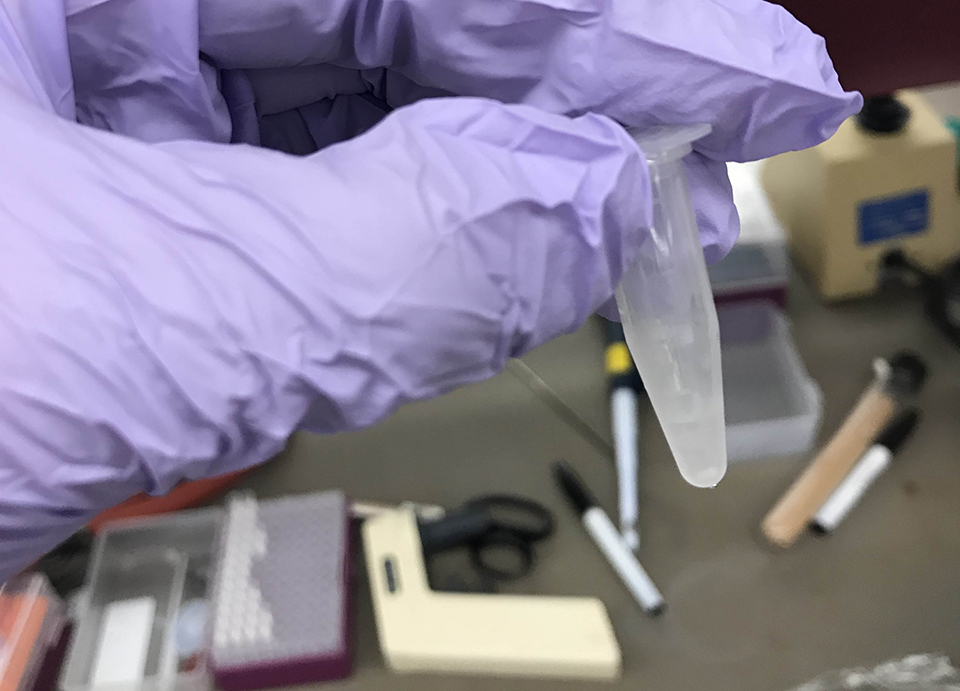
I. Acryloyl-X Treatment
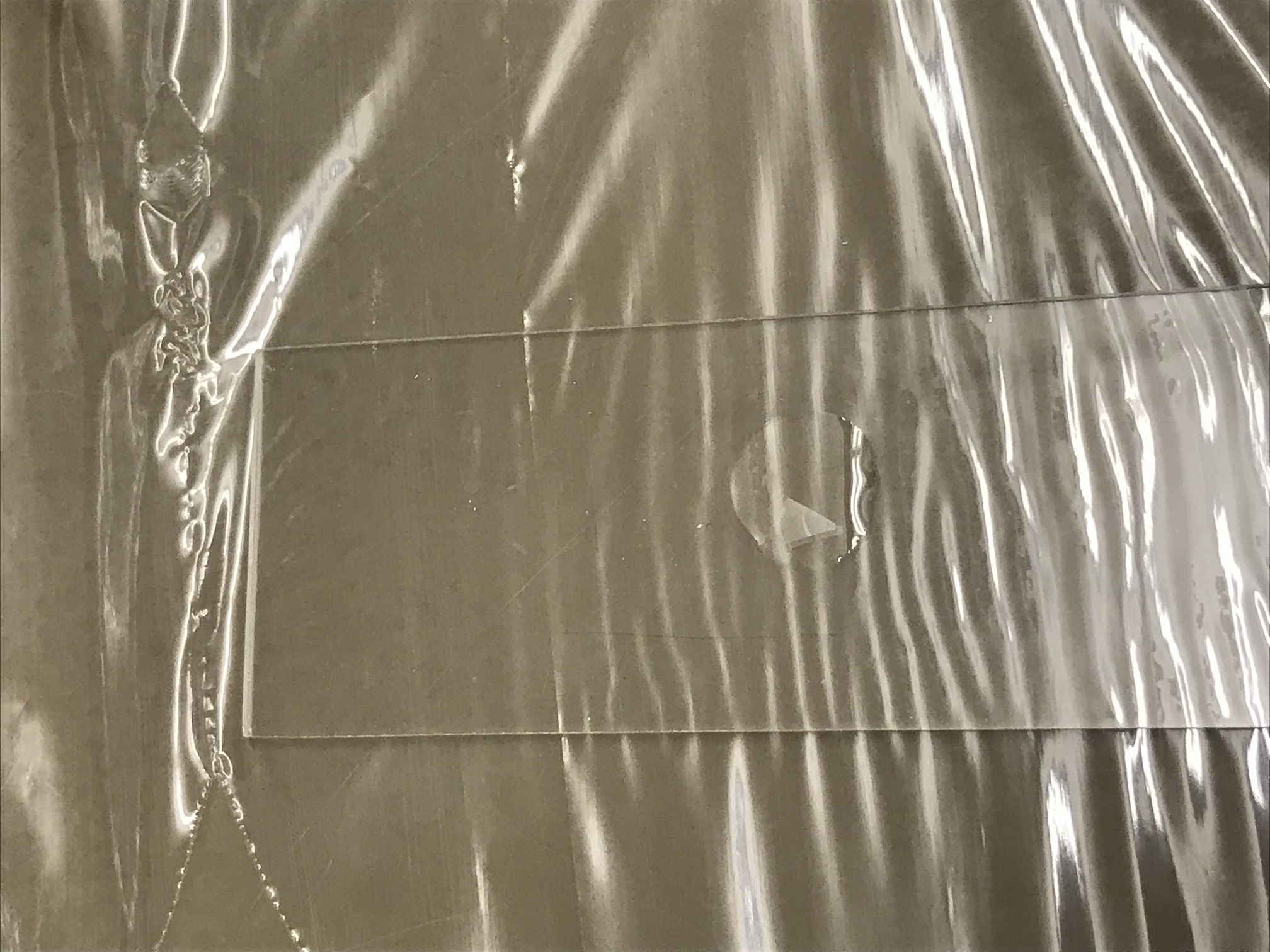
II. Gel-embedding and digestion
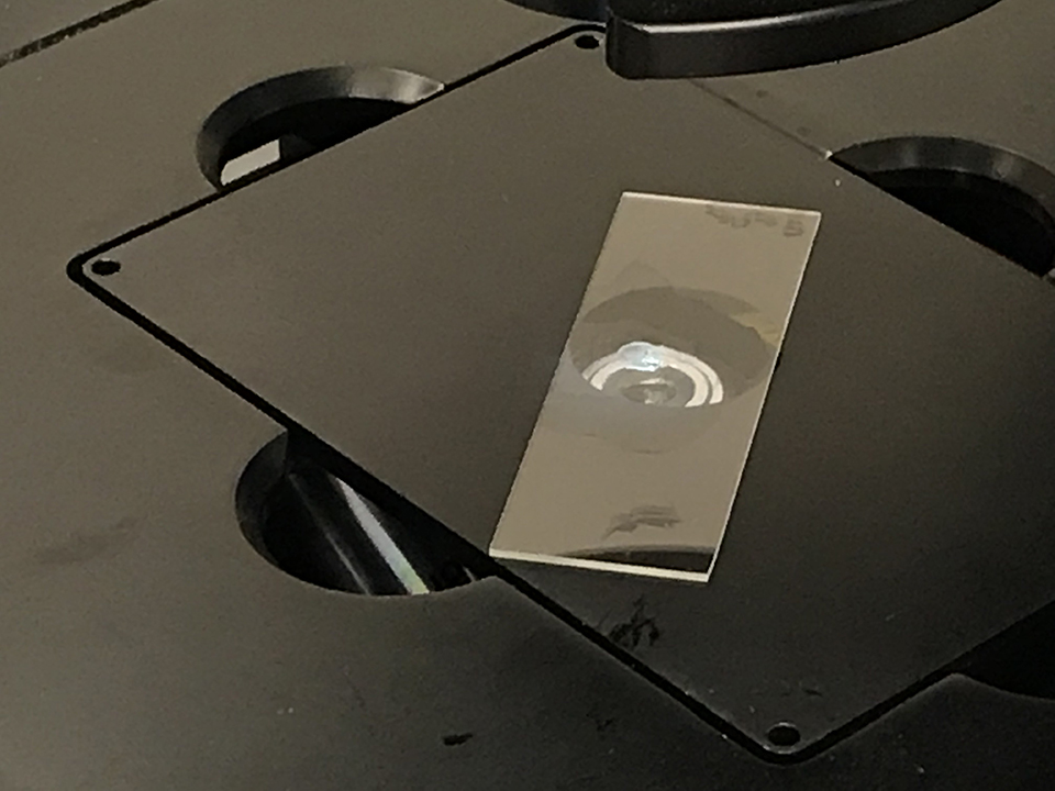
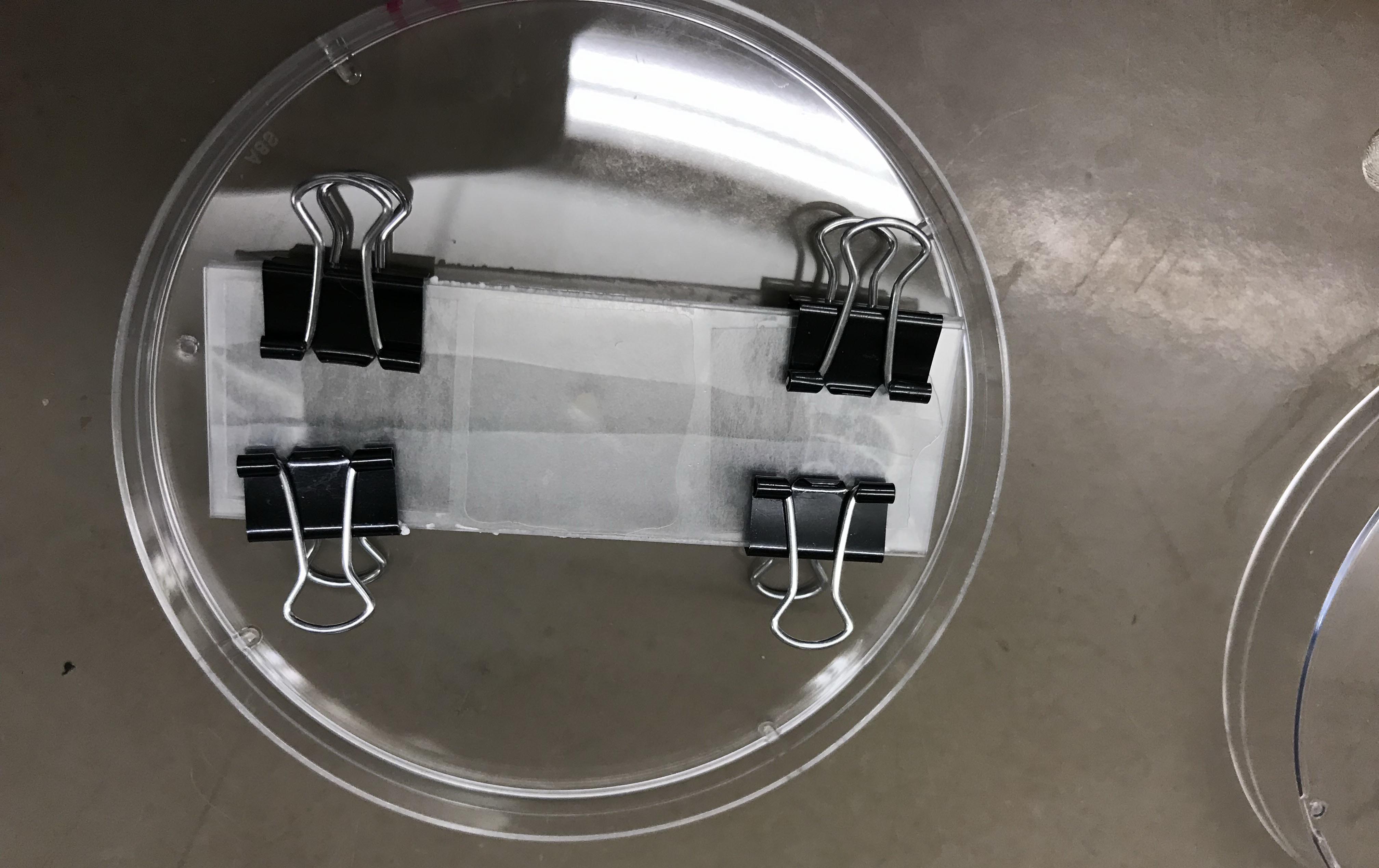
III. Expansion and visualization