Lab notes:
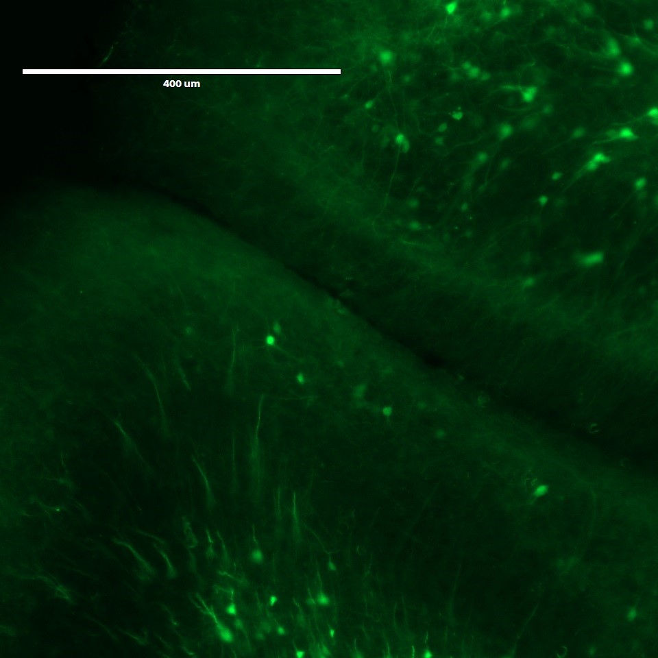
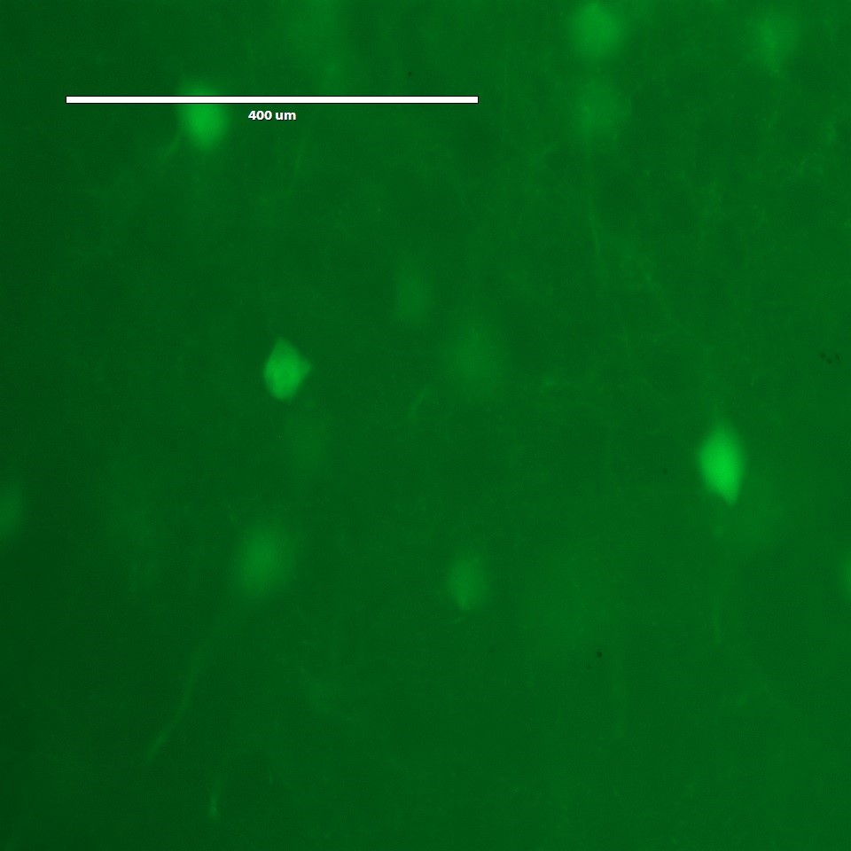
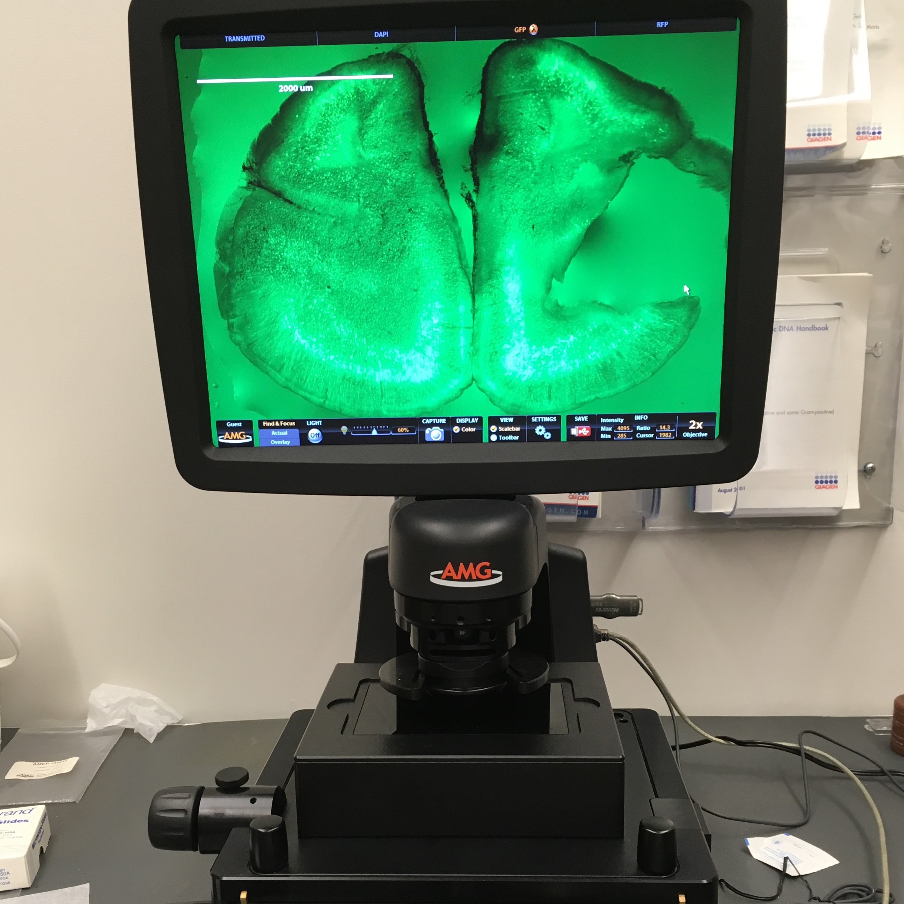
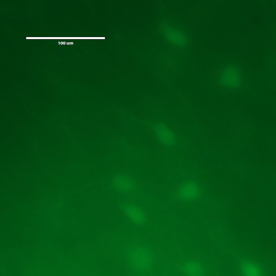
The image on the left is taken of the brain slice before we started the lab, and the image on the right is after the expansion. Looking at the size of the nuerons we can see it expanded A LOT!
This week prefromed an expanion microscopy experiment to scale up a mouse brain. This was truely amazing.
Expansion Microscopy (ExM): a sample preparation tool for biological samples that allows investigators to identify small structures by expanding them using a polymer system. This TED talk by Ed Boyden explains the inspiration, technique and use.
Central dogma of molecular biology: the two-step process, transcription and translation, by which the information in genes flows into proteins: DNA → RNA → protein. Transcription is the synthesis of an RNA copy of a segment of DNA. RNA is synthesized by the enzyme RNA polymerase.
Histone code: a hypothesis that the transcription of genetic information encoded in DNA is in part regulated by chemical modifications to histone proteins, primarily on their unstructured ends. Together with similar modifications such as DNA methylation it is part of the epigenetic code.
The image on the left is taken of the brain slice before we started the lab, and the image on the right is after the expansion. Looking at the size of the nuerons we can see it expanded A LOT!
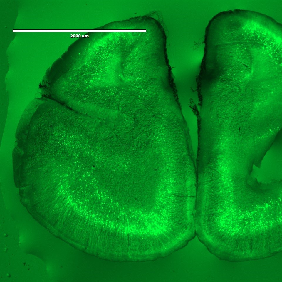
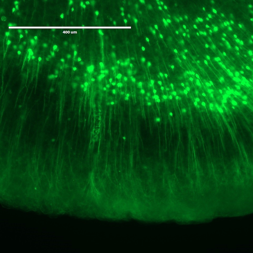
We took a slice of mouse brain and transfered it to a slide for imaging using a paiint brush. The brain tissue is very delicate and tore easily, so we had to be gentle and keep it moise with PBS. The initial images before expansion can be seen below at different magnifications.
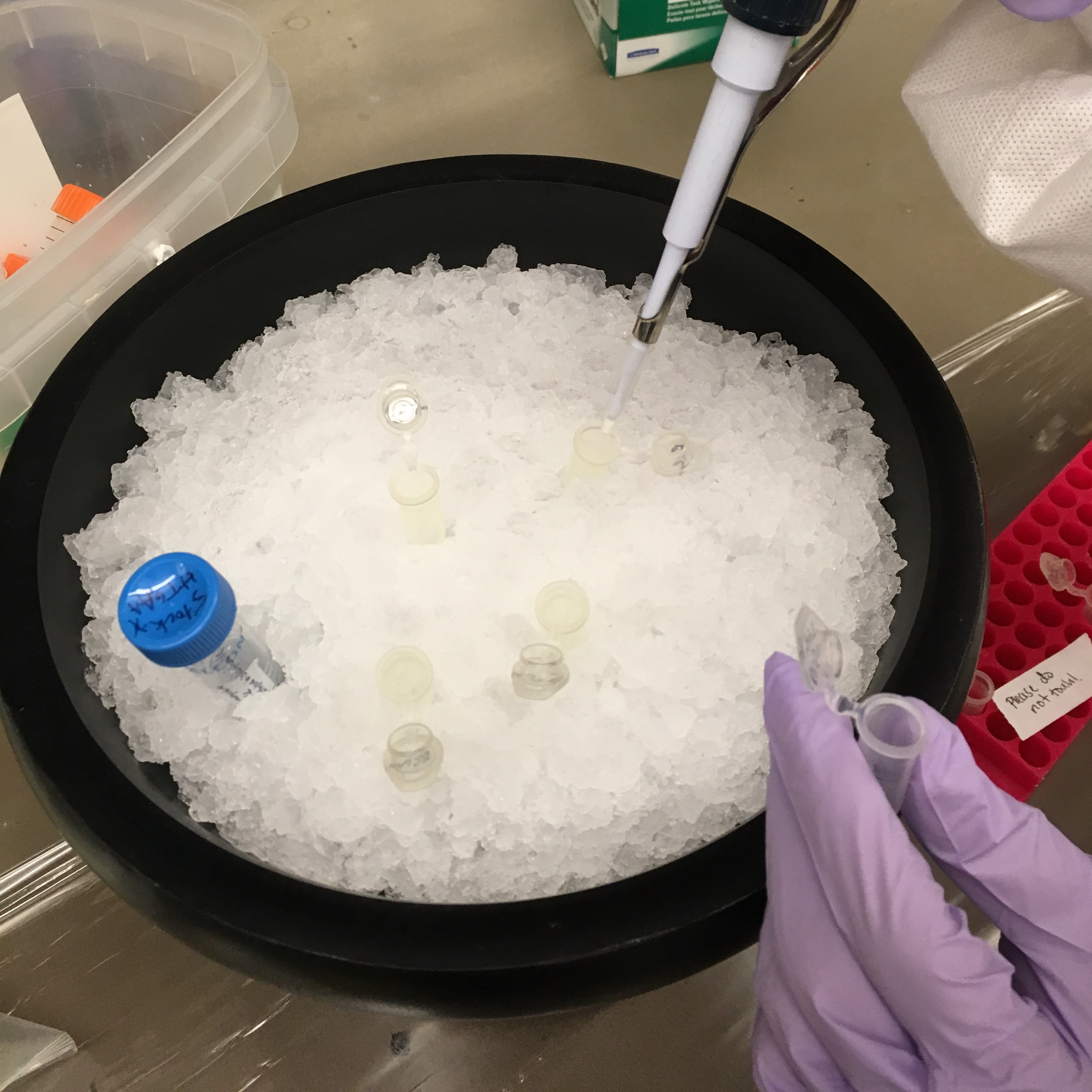
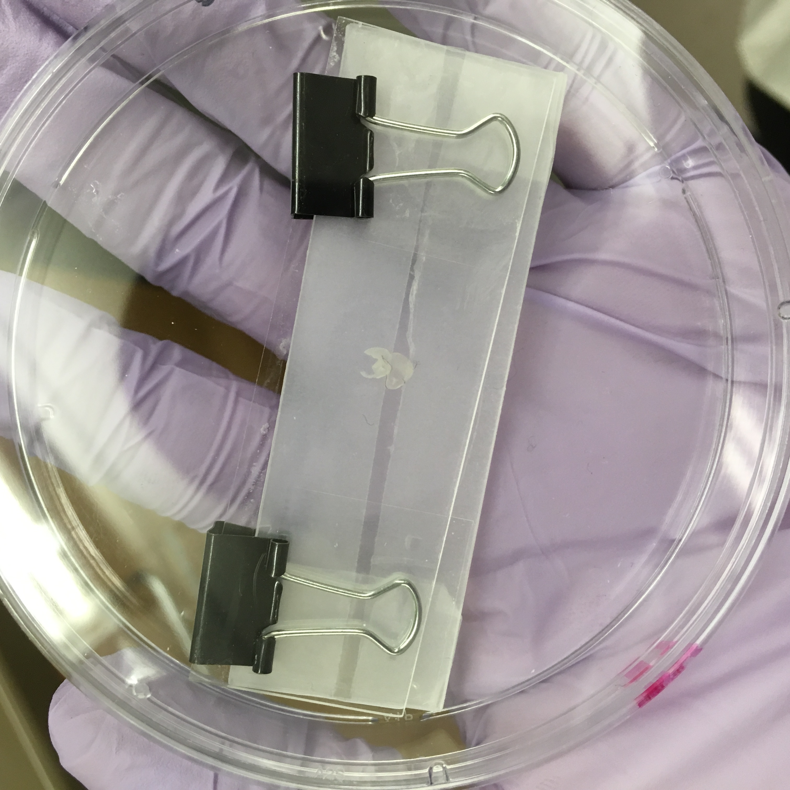
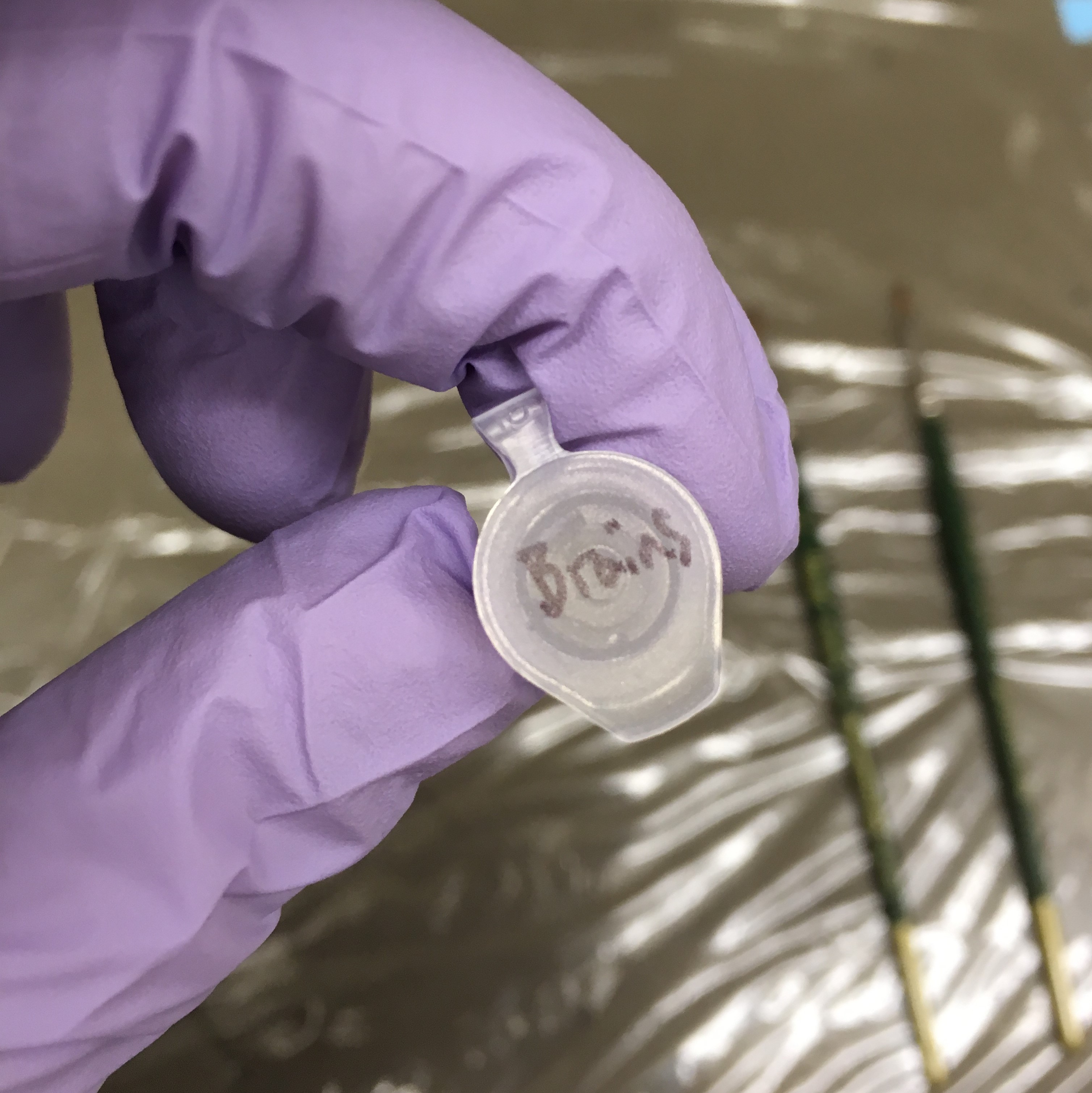
We prepaired the monomer solution following the lab procedure here. Since all the gel chemicals are toxic we covered the bench with saran wrap and all materials touching the gel were disposed of specially.
After soaking the brain slice in the gel for 30min, we put it in fresh monomer gel and sandwhiched it between slides separated by cover slips. We let this solidify for an hour then cut away the excess gel and put the sample into a 35mm petri dish with a digestion solution for expansion.