Week 10: Expansion Microscopy
Embiggening
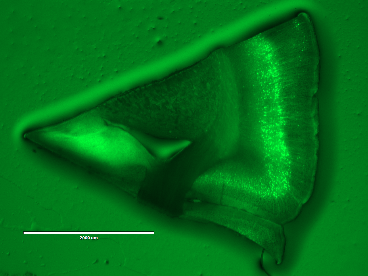
A brain slice before expansion
The idea of this module is to use gel expansion to clearly visualize YFP-labeled neurons in a slice of mouse brain. First we looked at the brain before expansion:
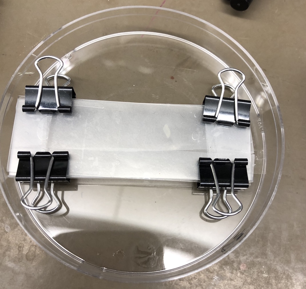
Making the gel
After putting the brain slice through successive rounds of toxic, gel-forming solution, we sandwiched it into a gel chamber surrounded by gel solution:
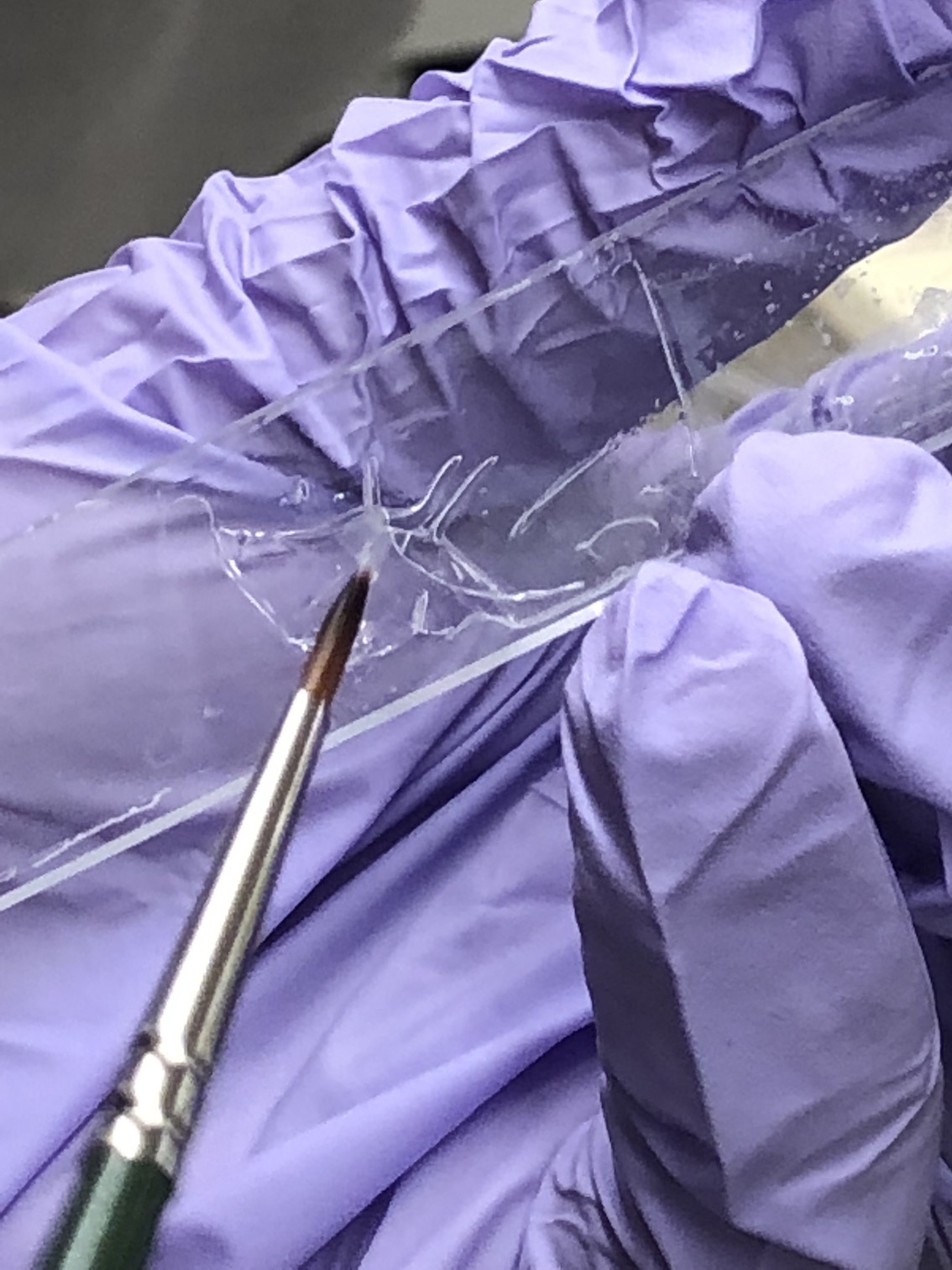
My sample was unsuccessful; no gel formed around the slice. Paul suggests that the parafilm wasn't stuck to the bottom glass slide, and thus rose and stuck to the top slide, pushing the gel out. The brain stuck to the parafilm, and we don't know why (parafilm is highly hydrophobic). Lucy's worked though, and she had to excise the brain slice from the gel sheet:
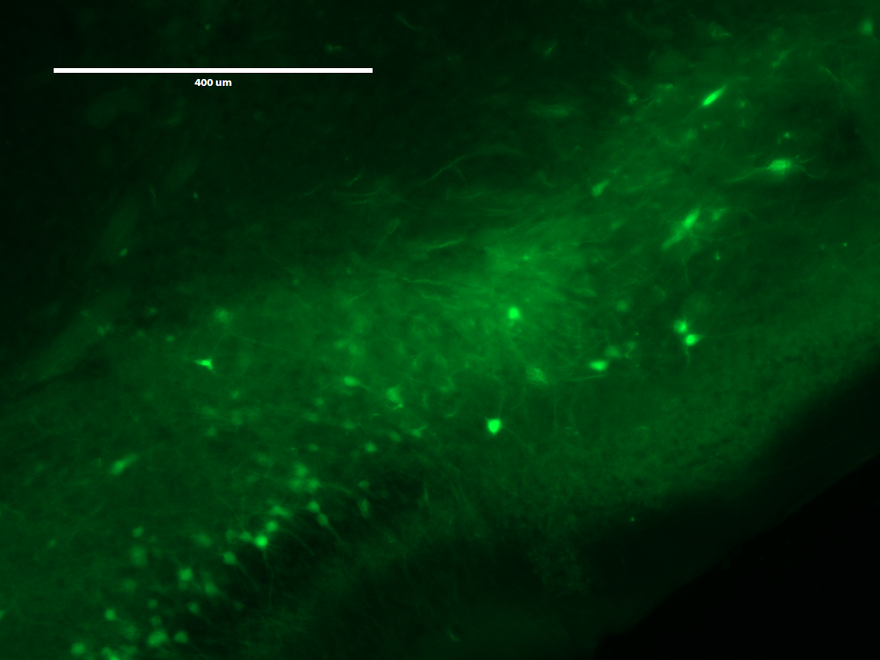
The expanded brain
After absorbing water overnight, Lucy's brain slice had expanded to nearly the size of the well! Visualizing under fluorescent microscope again, we can see individual neurons!
We used glass-bottom plates to enable the use of oil to increase resolution at 40x, but didn't get around to doing that.