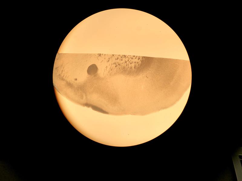
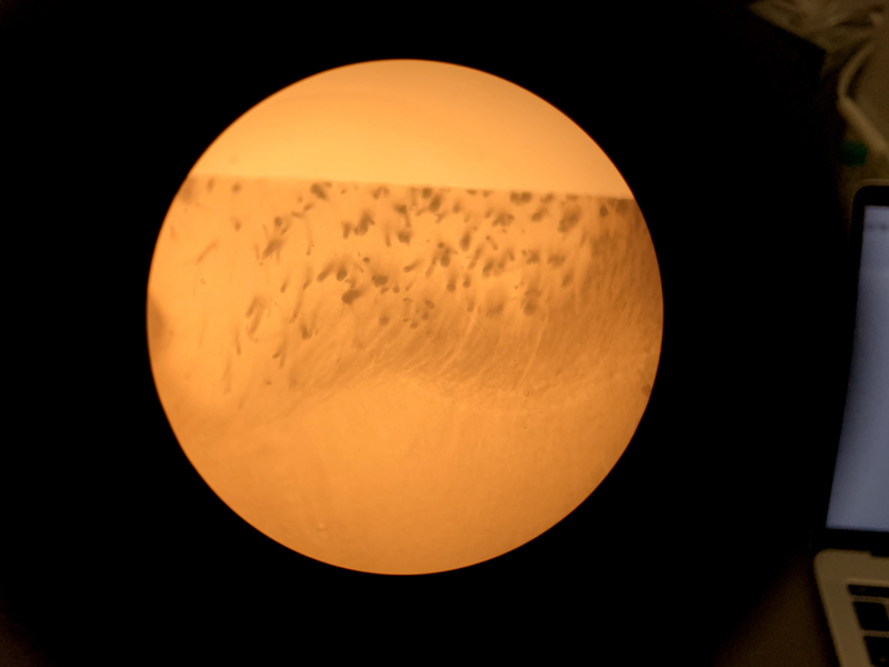
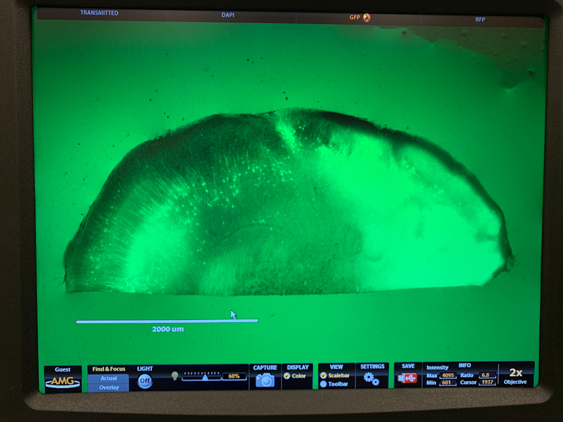
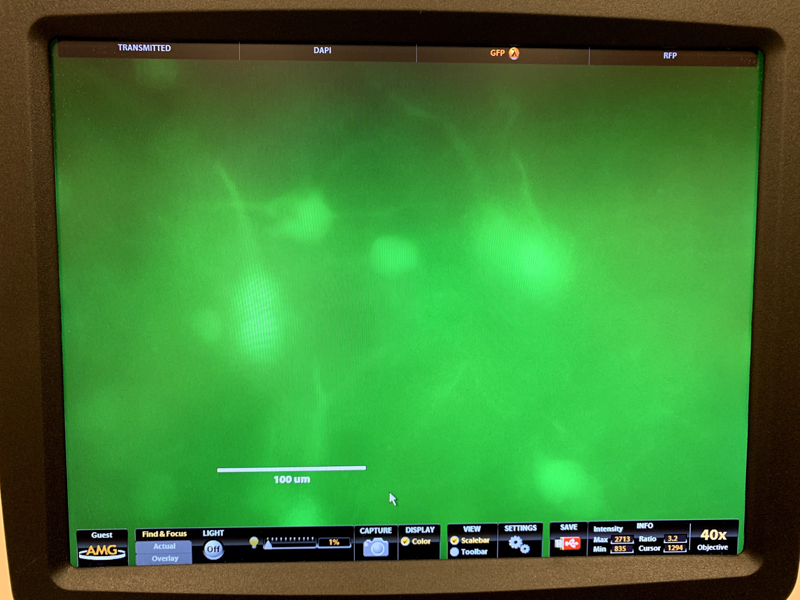
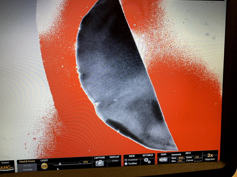
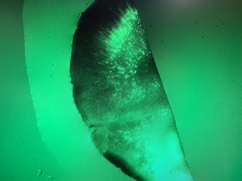
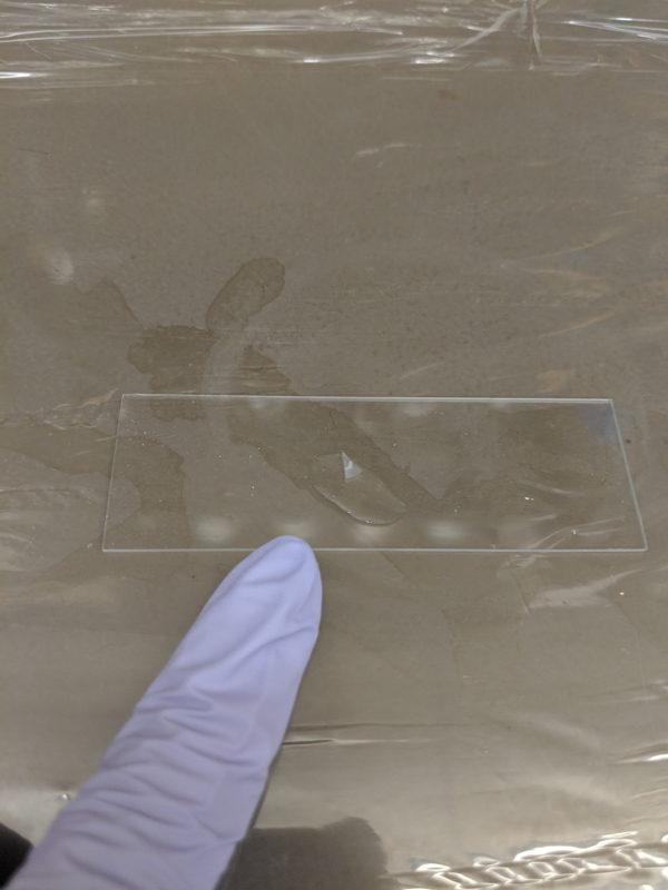
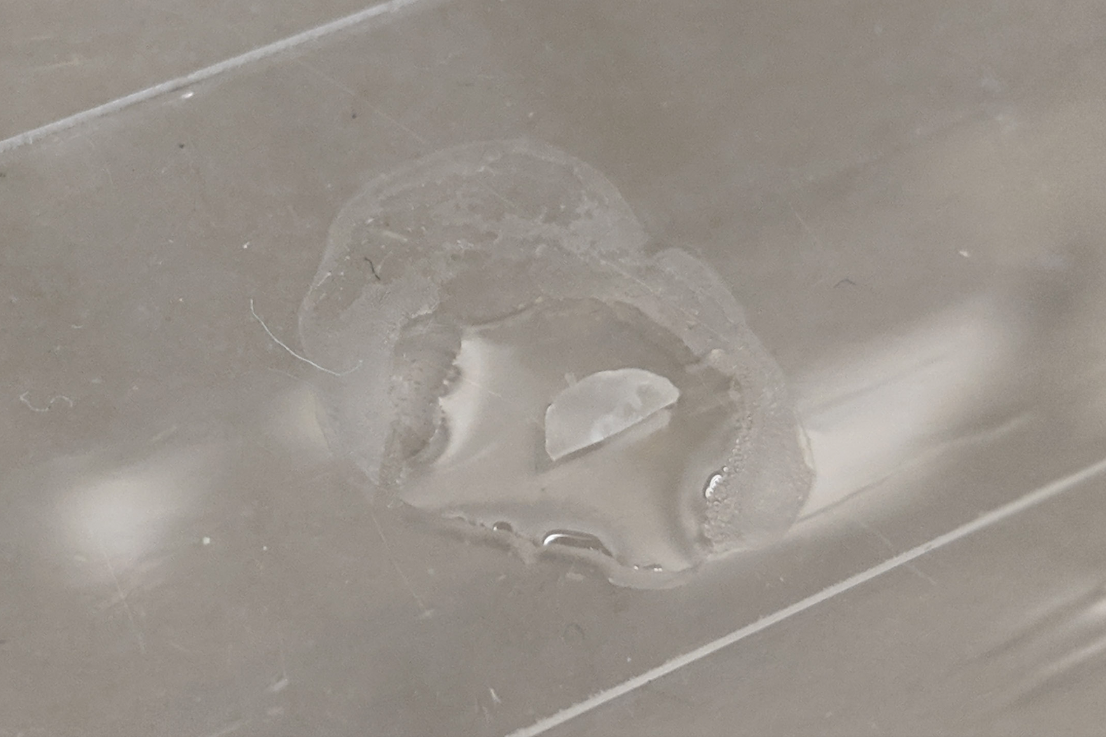
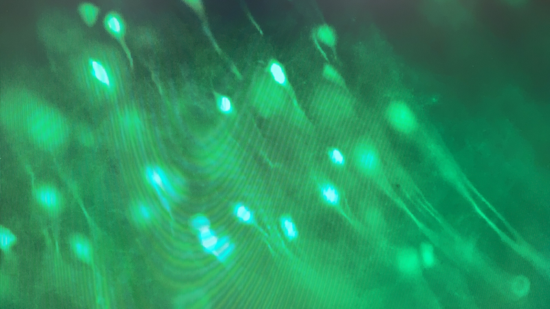
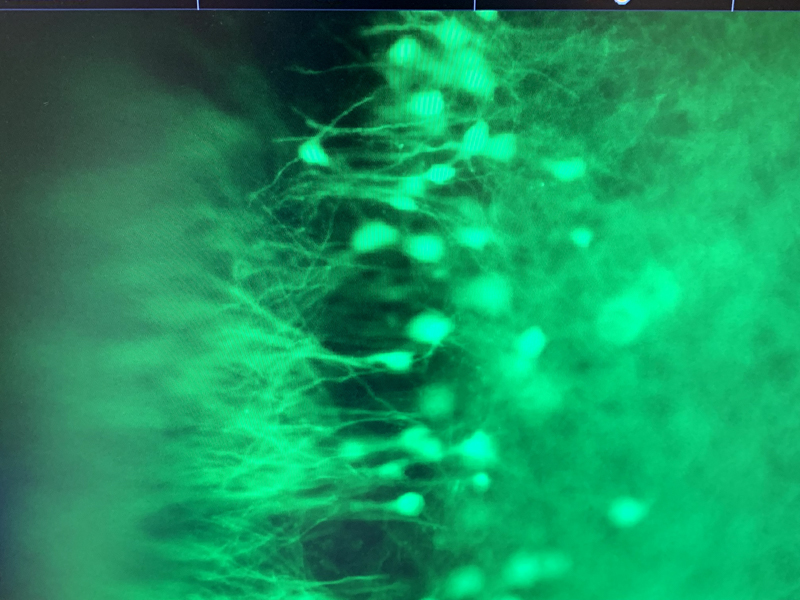
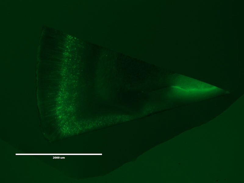
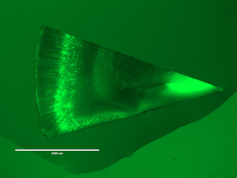
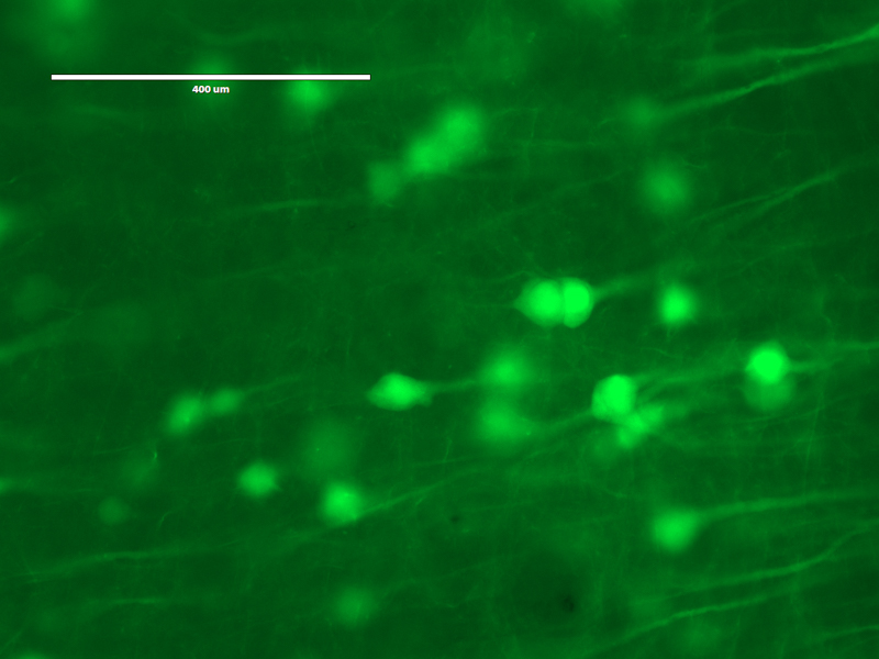
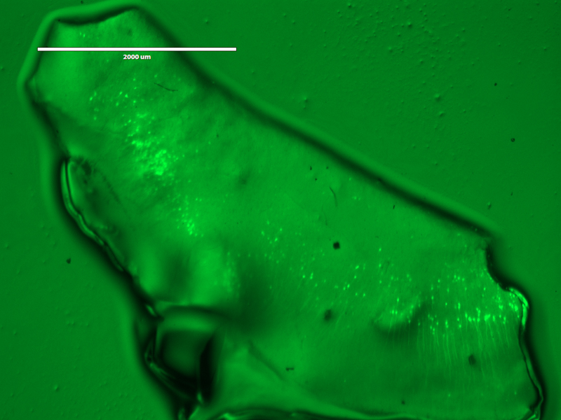
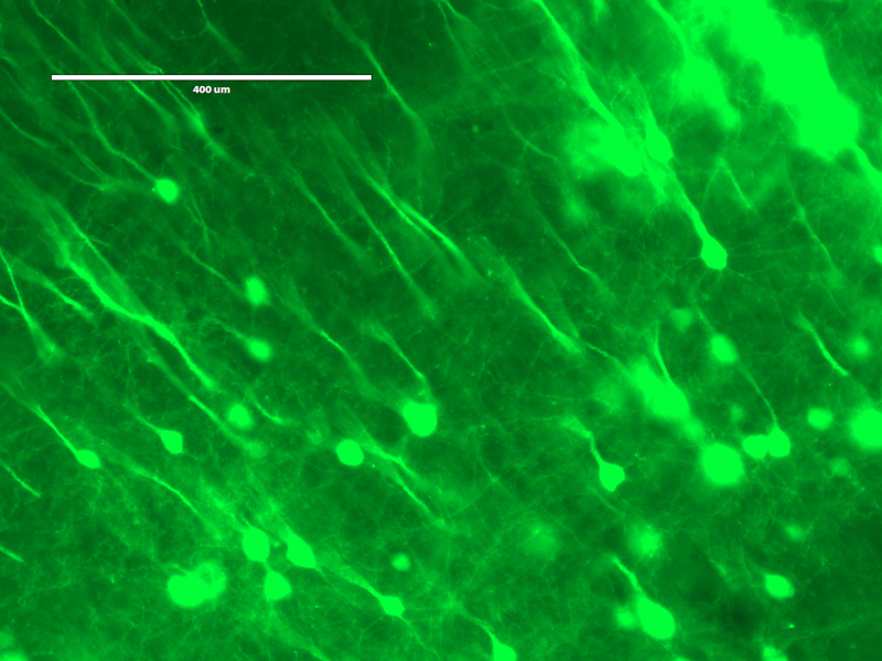
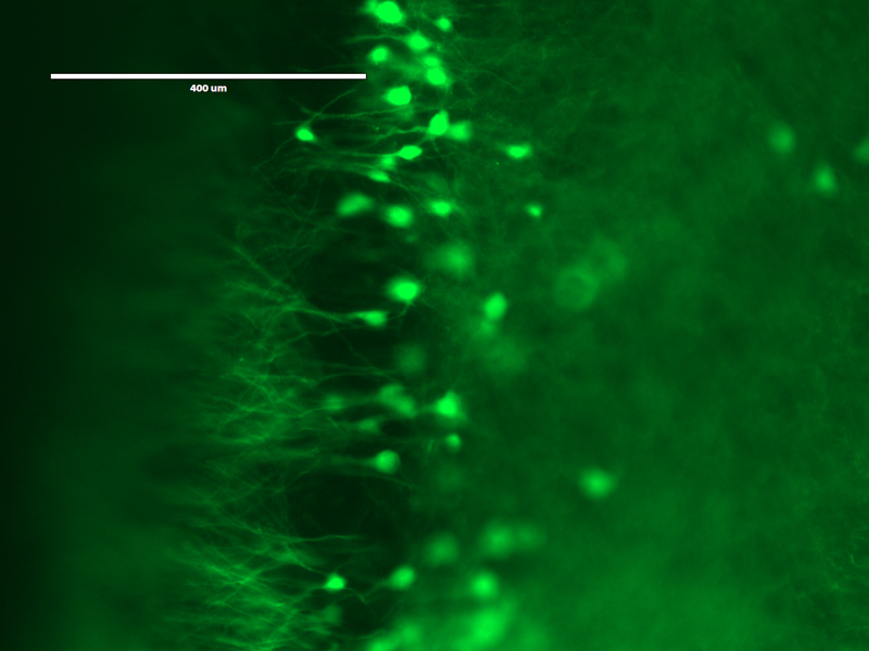
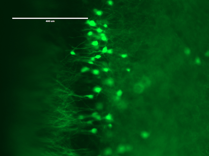
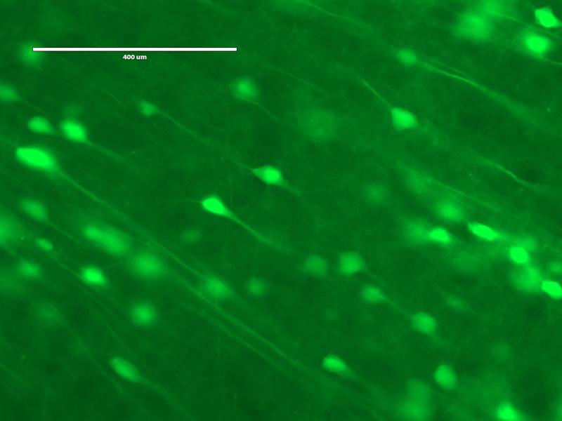
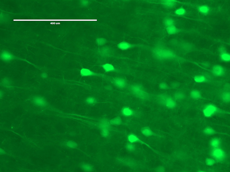
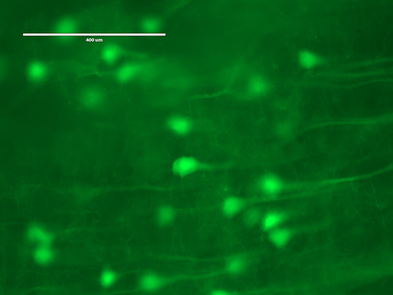
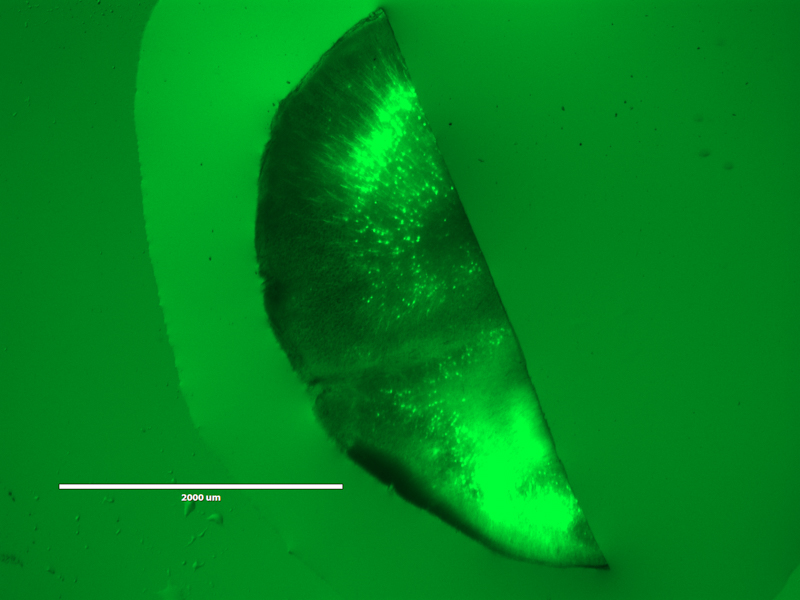
Use Expansion Microscopy (ExM) to to visualize a mouse brain slice with improved resolution on a light microscope. The tissue slices originate from a Thy1-YFP mouse, which expresses YFP in a subset of its neurons and thus permits visualization of neural structures. We will be performing protein-retension expansion microscopy (ProExM), wherein proteins in the sample are covalently linked to the gel before expansion. This variant of ExM is used to anchor fluorescent antibodies or fluorescent proteins directly to the gel. See the supplementary readings for more information about expansion microscopy and super-resolution imaging. For the published version of the protocol we’ll be implementing, see Protein-Retention Expansion Microscopy of Cells and Tissues Labeled Using Standard Fluorescent Proteins and Antibodies. Tillberg et al., Nature Biotechnology, 2016.